 | . | Step 28 of 41 |
 |
Clinical Stem 3
A patient with right upper lobe adenocarcinoma progressing on biomarker-directed therapy
A patient with right upper lobe adenocarcinoma progressing on biomarker-directed therapy
Answer B
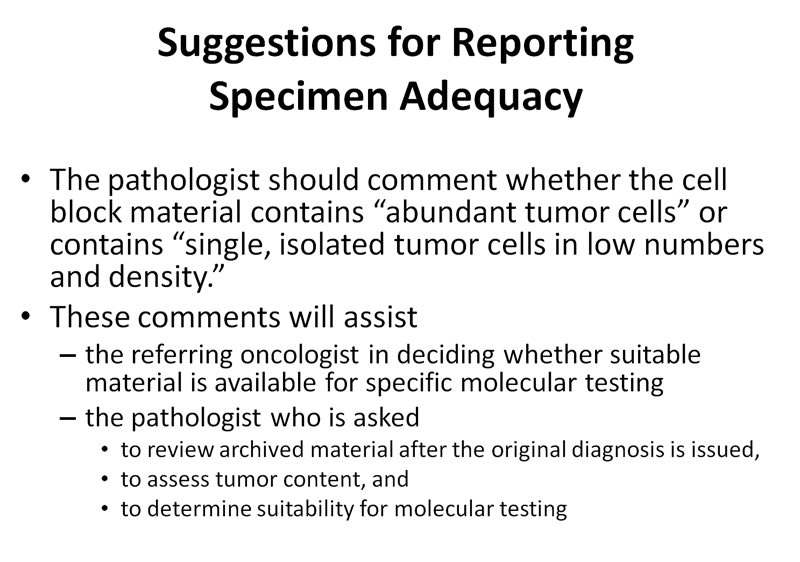
When cell blocks are planned, only a few slides might be needed for Diff-Quick and Papanicolau smear. The rest of the specimen is saved for cell-block preparation. Material obtained from additional sampling of the lesion or lymph node is expulsed from the needle into a solution such as Cytolyt. This is rendered into a pellet which is embedded in paraffin. This preparation may not represent the underlying tissue architecture, but is adequate for special stains and molecular analysis. Because there is some debate whether alcohol fixation is appropriate for FISH testing, techniques should be discussed with the institution's pathologist.

EBUS-TBNA and pleural fluid are more suitable than BAL because a larger number of tumor cells are usually retrieved, and there is greater possibility to make a cell-block.
Molecular testing for EGFR, K-RAS, BRAF, ALK and PIK3CA on cytology specimens provides results that are equivalent to those obtained from histology specimens in about 80% of cases.
Molecular testing for EGFR, K-RAS, BRAF, ALK and PIK3CA on cytology specimens provides results that are equivalent to those obtained from histology specimens in about 80% of cases.
Click here to download supplement materials
References:
- Burlingame OO, Kesse KO, Silverman SG, et al. On-site adequacy evaluations performed by cytotechnologists: correlation with final interpretations of 5241 image-guided fine needle aspiration biopsies. Cancer Cytopathol 2011.
- Colt HG, Murgu S. BronchAtlas. Smear preparation and bronchoscopic needle aspiration video available on YouTube (BronchOrg channel) 6/26/12.
- Muley T, Herth FJF, Schnabel P, Dienemann H, Meister M. From tissue to molecular phenotyping: Pre-analytical requirements Heidelberg Experience. Transl Lung Cancer Res 2012;1(2):111-121.
- Smouse JH, Cibas ES, Janne PA et al. EGFR mutations are detected comparably in cytologic and surgical pathology specimens of nonsmall cell lung cancer. Cancer 2009; 117: 67-72.
- Billah S, Stewart J, Staerkel G et al. EGFR and KRAS mutatiaons in lung carcinoma: molecular testing by using cytology specimens. Cancer Cytopathol 2011; 119:111-117.
- Schuurbiers OC, Looijen-Salamon MG, Liqtenberg MJ et al. A brief retrospective report on the feasibility of epidermal growth factor receptor and KRAS mutation analysis in transesophageal ultrasound and endobronchial ultrasound-guided fine needle cytological aspirates. J Thorac Oncol 2010; 5: 1664-1667).
- Pusztaszeri M, Soccal PM, Mach Net al. Cytopathological diagnosis of nonsmall cell lung cancer: recent advances including rapid on-site evaluation, novel endoscopic techniques and molecular test. J Pulmonar Respirat Med 2012; S5:002. Accessed June 12, 2013 http://dx.doi.org/10.4172/2161-105X.S5-002.


