 | . | Step 23 of 40 |
 |
Clinical Stem 2
A patient with pulmonary nodules 1 year after curative intent resection of primary lung adenocarcinoma
A patient with pulmonary nodules 1 year after curative intent resection of primary lung adenocarcinoma
Answer E
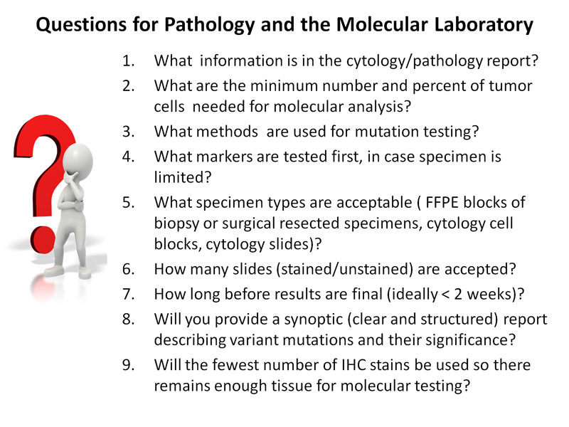
The pathologist should anticipate the appropriate use of IHC stains and molecular analysis to avoid wasting tissue unnecessarily for tests that are not required in the clinical situation.
Documenting the time of specimen acquisition is crucial for calculating time to specimen fixation. While fixation of lung cancer tissue has not been standardized, short fixation times of 6?12 hours for small biopsy specimens and 8?18 hours for larger resection specimens in 10% neutral buffered formalin are optimal for DNA and RNA-based tests, as well as FISH assays.
Samples should be examined by a pathologist to document the tumor's cellular content and purity in the area of tissue being sent for molecular analysis. Sample assessment is critical to obtain accurate results and to prevent false negatives.
An ideal sample has a high proportion of malignant cells relative to benign cells, and a low amount of substances such as mucin or necrotic tissue that may inhibit amplification.

Preserving scant tissue for most relevant tests is a major challenge facing pathologists who handle small volume cytology and histology specimens
Sending specimens directly to the molecular laboratory without prior assessment of tumor content by a pathologist should be avoided.
Sending specimens directly to the molecular laboratory without prior assessment of tumor content by a pathologist should be avoided.
Click here to download supplement materials
References:
- Thunnissen E, Kerr KM, Herth FJ, et al. The challenge of NSCLC diagnosis and predictive analysis on small samples. Practical approach of a working group. Lung Cancer 2012; 76: 1-18.
- Williams C, Ponten F, Moberg C, et al. A high frequency of sequence alterations is due to formalin fixation of archival specimens. Am J Pathol. 1999; 155:1467?1471.
- College of American Pathologists, International Association for the Study of Lung Cancer, Association for Molecular Pathology. Lung cancer biomarkers guideline draft recommendations. lung_public_comment supporting_materials.pdf. Published 2011. Accessed July 29, 2012.


